“The neurophysiological brain-fingerprint of Parkinson’s disease.”
Jason da Silva Castanheira, et al. – McGill University.
Background: Cortical neural activity gives rise to an individualized “brain-fingerprint.” Approaches using functional magnetic resonance imaging (fMRI) in patients with PD have found that brain-fingerprints are less stable than in healthy controls.
This Study: Castanheira and colleagues generated brain-fingerprints using magnetoencephalography (MEG), which captures neural activity on a finer time scale than fMRI, in patients with PD and healthy controls.
- Unlike in fMRI-based approaches, MEG-based brain-fingerprints could distinguish patients with PD from other patients, as well as classify subjects as patients or controls.
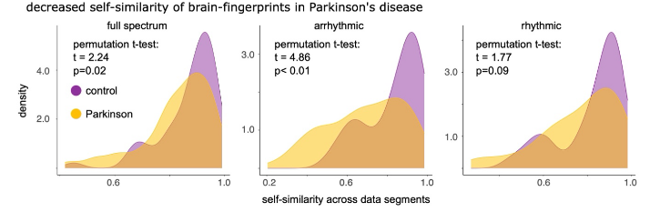
- Instability in patient brain-fingerprints was driven by the arrhythmic component, with the rhythmic component showing stability comparable to that seen in controls.
- Patients with later-stage PD showed a reduction in brain activity in the >15 Hz range and increased slow-wave (6-9 Hz) activity.
Bottom Line: MEG offers the means to study the individual cortical brain wave patterns of patients with PD.
Open Question: Could stimulation approaches that boost faster-wave activity and curb slower-wave activity provide therapeutic benefits?