Qi Wang, et al. – Sun Yat-Sen University.
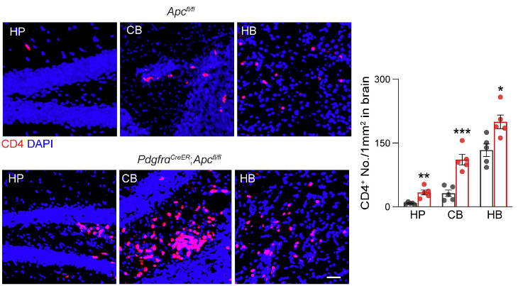
This study identified an ability for Wnt-expressing oligodendrocyte progenitor cells (OPCs) to enhance the autoimmune response in an MS model. Although OPCs in patients with MS express high levels of Wnts, this is not observed in mouse models of MS such as experimental autoimmune encephalomyelitis (EAE). After amplifying Wnt signaling in OPCs of mice subjected to EAE, Wang and collegaues demonstrated increased infiltration of Th1 cells into the brain, local transformation of macrophages into a cytotoxic profile, and exacerbated symptoms compared to standard EAE protocols. This effect is due to Wnt-driven OPC secretion of CCL4. Activated macrophages, in turn, release factors such as IL-18 that further increase Wnt expression in OPCs and lead to OPC death.
Since EAE symptoms are worse and closer to those observed in human MS when mouse OPCs are driven to express Wnt signals, this otherwise-absent element of immune system activation may help explain why many treatments successful at curbing EAE do not succeed in the clinic.