Manci Li and Peter Larsen – University of Minnesota.
Background: Neurons expressing high levels of neuropeptides (NPs; HNP neurons) are disproportionately lost in the entorhinal cortex (EC) during AD, but the mechanisms behind this vulnerability and whether it exists for HNP neurons in other regions remain unknown.
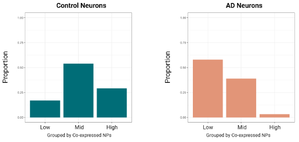
This Study: Li and Larsen first build on their prior work examining HNP neurons in the EC by comparing neurons expressing high and low levels of NPs in healthy controls, finding that HNP neurons are express higher levels of genes related to metabolism as well as innate immune signaling. Comparing neurons with medium NP loads between AD and controls, since HNPs are largely absent in AD, found that those in AD show reduced levels of genes associated with control HNPs, while upregulating genes related to pathways such as misfolded proteins and impaired mitochondrial signaling. Analysis of NP expression during healthy aging found that decreases were limited to regions affected by AD (with other regions such as the basal ganglia showing no changes) and to NPs linked to AD (ADNPs, with other NPs showing no changes in AD-linked brain regions). These effects are, at least in part, due to an enrichment for ADNP-expressing neurons in AD-linked brain regions. While the majority of these neurons are GABAergic interneurons, in line with expectations, around a quarter do not fit into any known subtype and express glutamatergic markers. Subjects with mild cognitive impairment had a distribution of NP-expressing neurons that resembled an intermediary between the control and AD profiles, suggesting that the selective loss of HNP neurons is a driver of progression from typical cognition to MCI and further on to AD.
Bottom Line: The vulnerability of HNP neurons and their enrichment in areas such as EC may represent key factors in the development and progression of AD. Previous studies describing the vulnerability of many of these neuronal populations have largely viewed them through the lens of being GABAergic interneurons, focusing on how the loss of their inhibitory input could promote the observed hyperactivity of excitatory neurons in AD. These results suggest that their role as HNP neurons should not be ignored, particularly given that some of these neurons are not GABAergic.