Léa Chauveau, et al. – Normandy University.

Background: Changes to the medial temporal lobe are critical elements of AD pathology. However, there are conflicting data about how networks within this region are affected during AD. In addition, it remains unknown how these changes relate to markers of AD pathology and to what extent these changes could be attributed to aging.
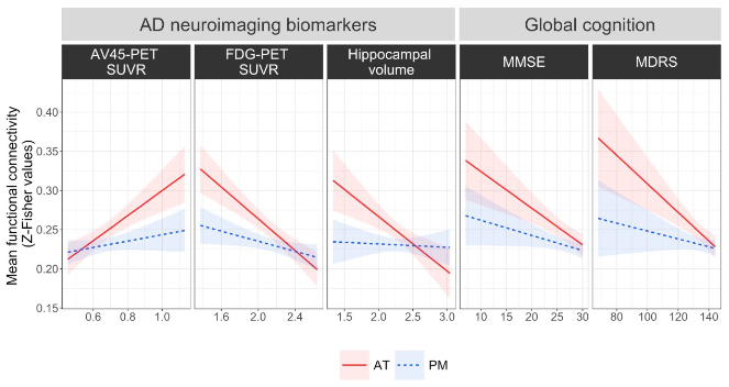
This Study: Using longitudinal data from adults ages 19 to 85 (n=267), with older adults spanning the spectrum from cognitively unimpaired (CU, n=56) to mild cognitive impairment (MCI, n=26) and AD (n=26), Chauveau and colleagues found that connectivity within the anterior-temporal (AT) network positively correlated with both aging (young vs old) and cognitive decline (CU vs MCI vs AD, all older adults). Increased AT also correlated with worse AD neuroimaging and cognitive assessments, as well as faster time to convert from MCI to AD among subjects who did so during the study period. In contrast, connectivity within the posterior-medial (PM) network decreased with normal aging and had no association with AD status.
Bottom Line: Connectivity within the AT network may represent an imaging biomarker for AD; better understanding of why connectivity increases in AD could lead to therapeutic treatments to combat it.


