Hao Chen, et al. – Weill Cornell Medicine.
Background: Previous studies found Dap12 knock-out (KO) reduces memory deficits and synaptic impairments in various Alzheimer’s disease (AD) mouse models, despite also increasing tau pathology.
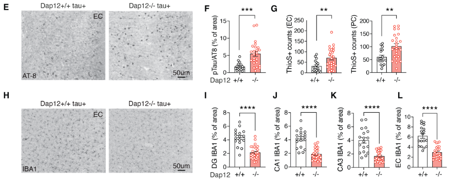
This Study: Chen and colleagues studied transcriptomic changes to microglia and oligodendrocytes (OLs) induced by Dap12 KO in the Tau P301S mouse model, finding a broad shift away from disease-associated states. These include reduced cytokine expression in microglia and increased myelination within the dentate gyrus. One tau-associated OL subtype was reduced in number following Dap12 KO, and this subtype was more prevalent in brain samples from patients with AD compared to controls of similar age, tau burden, and clinical dementia ratings, suggesting this subtype is involved in AD-specific mechanisms. They also observed lower levels of synapse loss in the CA1 region of the hippocampus, in line with prior studies finding Dap12 KO reversed tau-associated memory deficits in the Barnes water maze and Novel Object Recognition assays.
Bottom Line: DAP12 is a critical component of the microglial signaling pathways tying tau accumulation to neuroinflammatory signaling.